The cardiac conduction system is the electrical pathway of the heart that includes in order the SA node AV node bundle of His bundle branches and Purkinje fibers. Cell theory has its origins in seventeenth century microscopy observations but it was nearly two hundred years before a complete cell membrane theory was developed to explain what separates cells from the outside world. Cell membrane diagram labeled simple.
Cell Membrane Diagram Labeled Simple, The cell membrane provides mechanical support that facilities the shape of the cell while enclosing the cell and its components from the external environment. That brief rise to 50 mV at point A on the axon however causes the potential to rise at point B leading to an ion transfer there causing the potential there to shoot up to 50 mV thereby affecting the potential at point C etc. Studies of the action of anesthetic molecules led to. MATHEMATICAL MODEL The Hodgkin-Huxley model is based on the parallel thought of a simple circuit with batteries resistors and capacitors.
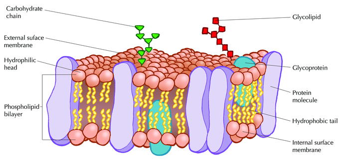 The Fluid Mosaic Model Introducing The Cell From nigerianscholars.com
A basic model of this circuit is shown in Figure 4. Easily learn the conduction system of the heart using this step-by-step labeled diagram. Explore the definition function and structure of the. Membrane-bound Arf-GDP which is important for GBF1 recruitment is probably also not the limiting factor because total membrane-bound Arf increases significantly after Src activation.
The first is a colored and labeled cell diagram.
Read another article:
Learn about pacemaker cells and cardiac ac. The first is a colored and labeled cell diagram. Use this convenient study aid in preparation for your upcoming test or quiz. This enhanced visual instructional tool assists in grasping and retaining the names of the cell parts like mitochondrion vacuole nucleus and more with ease. CPR5 PNET1 GP210 and NDC1 are structural components of the plant NPC membrane ring.
 Source: study.com
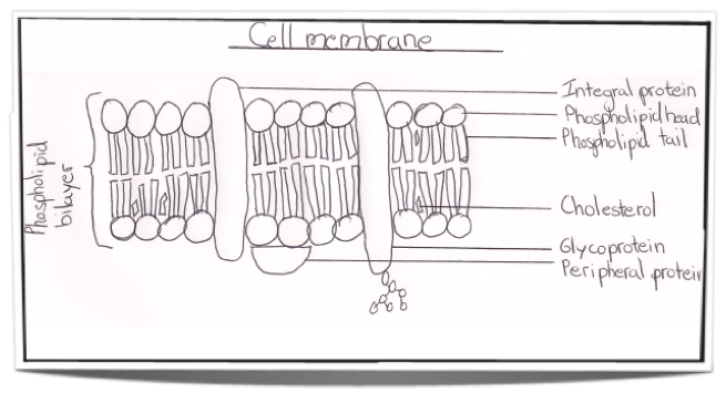
If so you may need to memorize the animal cell its organelles and their functions. Because it does not have a hard cell wall animal cells vary in shape. Structures Unique to Plant Cells. The limiting factor may arise from the export of GALNTs-containing carriers resulting in the depletion of an unidentified receptor on Golgi membranes. Draw And Label A Simple Line Diagram Of A Cell Membrane Include The Major Types Of Membrane Proteins On Your Diagram Also Indicate How The Following Pass Through The Cell Membrane Oxygen.
 Source: ibguides.com
The simple epithelial tissue is a closed network of flat epithelial cells. It regulates what can be allowed to enter and exit the cell through channels acting as a semi-permeable membrane which facilities the exchange of essential compounds required for the survival of the cell. The animal cell is made up of several structural organelles enclosed in the plasma membrane that enable it to function properly eliciting mechanisms that benefit the host animal. Signaling molecules can belong to several chemical classes. Ib Biology Notes 2 4 Membranes.
 Source: shutterstock.com
These are located on the basal membrane. It is composed of a single layer of cells that are specialized in diffusion osmosis filtration secretion and absorptionThe simple epithelial tissue is found in the alveolar epithelium pulmonary alveolus the endothelium lining of blood vessels and lymph. The cytoplasm enclosed within the cell membrane does not exhibit much structure when viewed by electron microscopy. This prevents the action potential from travelling backwards. Cell Membrane Images Stock Photos Vectors Shutterstock.
 Source: nigerianscholars.com
A biosensor is an analytical device containing an immobilized biological material enzyme antibody nucleic acid hormone organelle or whole cell which can specifically interact with an analyte and produce physical chemical or electrical signals that can be measured. Studies of the action of anesthetic molecules led to. A biosensor is an analytical device containing an immobilized biological material enzyme antibody nucleic acid hormone organelle or whole cell which can specifically interact with an analyte and produce physical chemical or electrical signals that can be measured. The animal cell is made up of several structural organelles enclosed in the plasma membrane that enable it to function properly eliciting mechanisms that benefit the host animal. The Fluid Mosaic Model Introducing The Cell.
 Source: drawitneat.blogspot.com
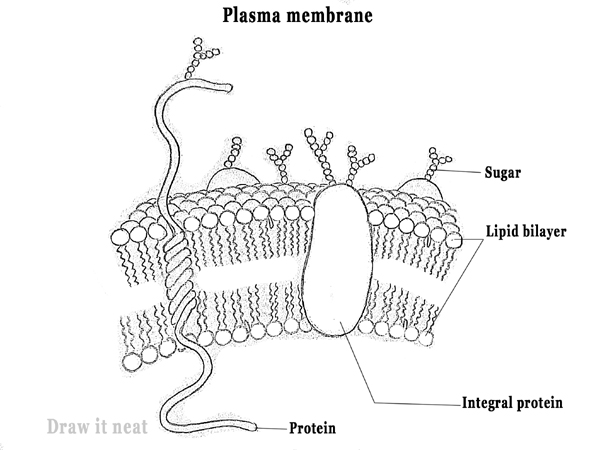
The cardiac conduction system is the electrical pathway of the heart that includes in order the SA node AV node bundle of His bundle branches and Purkinje fibers. Living cells are divided into two types - prokaryotic and eukaryotic sometimes spelled procaryotic and eucaryotic. A basic model of this circuit is shown in Figure 4. A membrane is a selective barrier which allows certain things to pass through but not othersfor this reason the cell membrane is said to be selectively permeable. Draw It Neat How To Draw Plasma Membrane Cell Membrane.
 Source: pinterest.com
The simple epithelial tissue is a closed network of flat epithelial cells. The limiting factor may arise from the export of GALNTs-containing carriers resulting in the depletion of an unidentified receptor on Golgi membranes. Easily learn the conduction system of the heart using this step-by-step labeled diagram. A biosensor is an analytical device containing an immobilized biological material enzyme antibody nucleic acid hormone organelle or whole cell which can specifically interact with an analyte and produce physical chemical or electrical signals that can be measured. Honors Biology Lawrenceville Cells Plasma Membrane Cell Membrane Cell Membrane Structure.
 Source: microbenotes.com
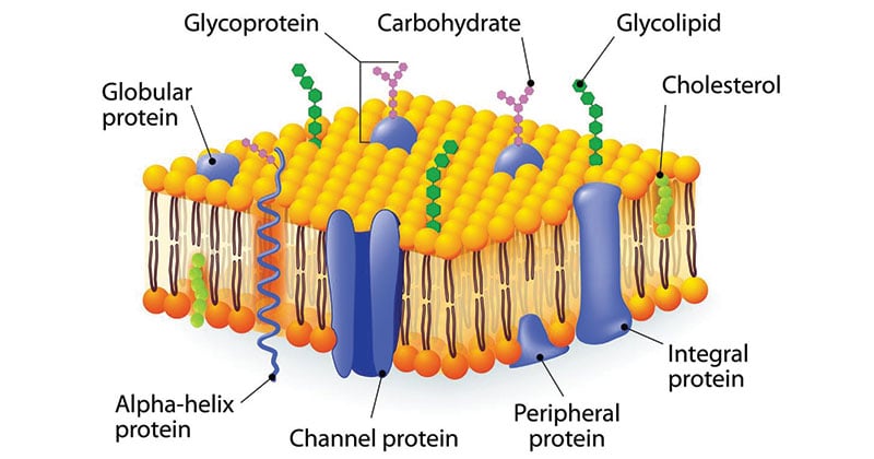
The animal cell is made up of several structural organelles enclosed in the plasma membrane that enable it to function properly eliciting mechanisms that benefit the host animal. Bacteria Prokaryotes are simple in structure with no recognizable organelles. While the membrane is hyperpolarized below resting the cell cannot fire. Featured in this printable worksheet are the diagrams of the plant and animal cells with parts labeled vividly. Membrane Carbohydrate Cell Biology Microbe Notes.

CPR5 PNET1 GP210 and NDC1 are structural components of the plant NPC membrane ring. Sequential centrifugation can be used to prepared endosomes. Learn vocabulary terms and more with flashcards games and other study tools. Bacteria Prokaryotes are simple in structure with no recognizable organelles. Plasma Membrane Teaching Resources.
 Source: dreamstime.com
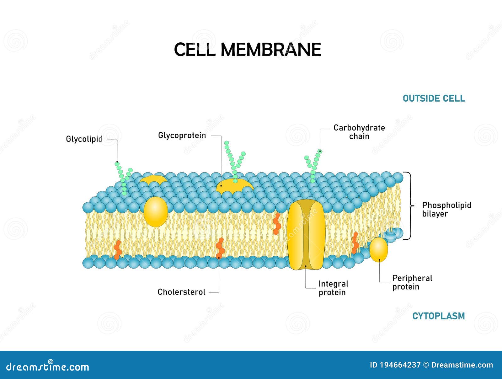
Hydrophobic membrane filters are typically used with compatible non-aqueous fluids. Hydrophobic membrane filters are typically used with compatible non-aqueous fluids. Explore the definition function and structure of the. At this point the membrane becomes impermeable to sodium again and potassium ions flow out of the cell restoring the axon at that point to its rest state. Diagram Of Cell Membrane Phospholipid Bilayers Structure Stock Vector Illustration Of Diffusion Biology 194664237.
 Source: www2.yvcc.edu
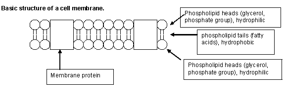
A biosensor is an analytical device containing an immobilized biological material enzyme antibody nucleic acid hormone organelle or whole cell which can specifically interact with an analyte and produce physical chemical or electrical signals that can be measured. The daughter cells are. The NE hosts a specific population of proteins. Cell Membrane Function In Animal Cell. Membrane Transport.
 Source: cronodon.com
This prevents the action potential from travelling backwards. The cell membrane also known as the plasma membrane is a barrier that surrounds a cell separating its interior from the outside environment. The cytoplasm enclosed within the cell membrane does not exhibit much structure when viewed by electron microscopy. To help you do this Ive created a printable animal cell diagram. Membranes.
 Source: dummies.com
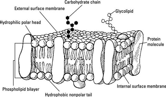
If so you may need to memorize the animal cell its organelles and their functions. They have an outer cell wall that gives them shape. A membrane is a selective barrier which allows certain things to pass through but not othersfor this reason the cell membrane is said to be selectively permeable. This enhanced visual instructional tool assists in grasping and retaining the names of the cell parts like mitochondrion vacuole nucleus and more with ease. The Cell Membrane Diffusion Osmosis And Active Transport Dummies.
 Source: shutterstock.com
It regulates what can be allowed to enter and exit the cell through channels acting as a semi-permeable membrane which facilities the exchange of essential compounds required for the survival of the cell. Chromosomes condense nuclear membrane dissolvesMitosis. That brief rise to 50 mV at point A on the axon however causes the potential to rise at point B leading to an ion transfer there causing the potential there to shoot up to 50 mV thereby affecting the potential at point C etc. SUN and KASH proteins comprise the LINC complex and function in various aspects of plant cell biology and physiology as discussed in the main text. Cell Membrane Images Stock Photos Vectors Shutterstock.
 Source: courses.lumenlearning.com
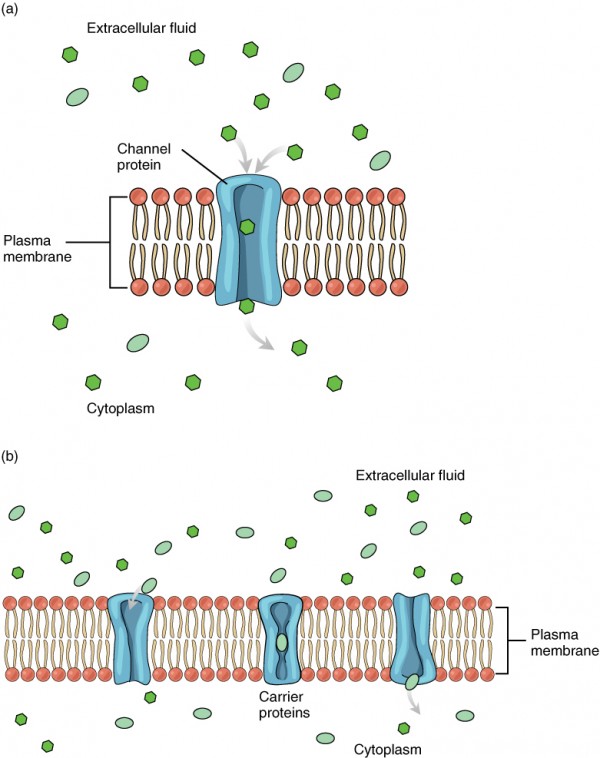
While the membrane is hyperpolarized below resting the cell cannot fire. Cell Membrane Function In Animal Cell. Use this convenient study aid in preparation for your upcoming test or quiz. Cell theory has its origins in seventeenth century microscopy observations but it was nearly two hundred years before a complete cell membrane theory was developed to explain what separates cells from the outside world. Membrane Transport Anatomy And Physiology.
 Source: biologycorner.com
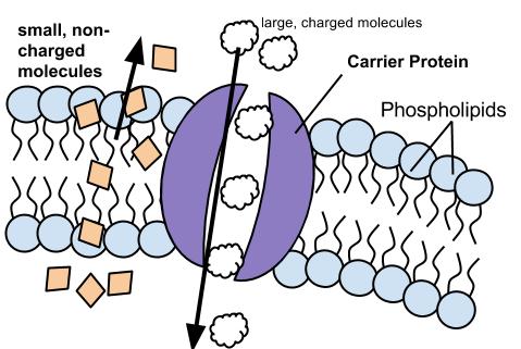
Easily learn the conduction system of the heart using this step-by-step labeled diagram. The NE hosts a specific population of proteins. The cardiac conduction system is the electrical pathway of the heart that includes in order the SA node AV node bundle of His bundle branches and Purkinje fibers. Easily learn the conduction system of the heart using this step-by-step labeled diagram. Ch 5 Membrane Structure And Function.


